Magnetic Resonance Imaging
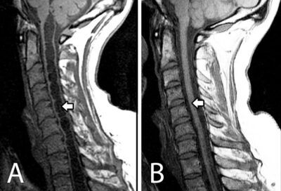
Magnetic resonance imaging (MRI) is currently the diagnostic test of choice for syringomyelia as it is able to detect fluid movement, syrinxes, and other abnormalities (Brodbelt & Stoodley 2003). MRI also has an important role in planning and monitoring outcomes of treatments. T1-weighted (T1W) and T2-weighted (T2W) images can be obtained from a MRI and allow for image contrast between different types of tissue (Enzmann 1991). Phase contrast with MRI can identify obstruction of the subarachnoid space and demonstrate normalization of CSF flow following surgery (as seen in Figure 2; Brodbelt & Stoodley 2003). However, MRI is limited in its ability to differentiate posttraumatic syringomyelia from myelomalacia or tumor-associated syringomyelia (Biyani & El Masry 1994).